Did you know there are about 100 different types of arthritis? However, we are going to focus on the most common form of arthritis called osteoarthritis (OA). Most everybody would consider this arthritis to be the “wear and tear” or “old age” arthritis. About 2% of the US population under the age of 45 suffers from OA, 30%suffers from OA between the ages of 45-64 and 65-85% suffers from OA over the age of 65. So, generally speaking this is a chronic disease that becomes more prevalent as one ages. It should be remembered that OA changes to the joint develop years earlier before significant pain is noted.
Based on the National Health Survey from 2007-2009, revealed 50 million or about 22% of adults have self reported doctor diagnosed OA. 21 million or about 9% have arthritis related reduction in activity. OA is the most common cause of disability particularly after age 50.
From the journal, Arthritis Rheumatology 2008, nearly 1 in 2 people will develop symptomatic knee OA by age 85 and 2 in 3 people who are obese ,which is a huge risk factor, may develop symptomatic knee OA in their lifetime. 1 in 4 may develop painful hip arthritis in their lifetime according to the journal Osteoarthritis and Cartilage.
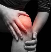
The most common sites that OA occurs are the knees, hips, spine and hands. Of course almost any other joint in the body can also be affected by OA. Due to it widespread presence about 25% of all visits to primary care physicians (PCP) are due to OA. Another interesting statistic is that 50% of those visits are due to complications from taking non-steroidal antiinflammatory drugs (NSAIDS) such as, aspirin, ibuprofen, naproxen, Celebrex, Voltaren and others.
In brief, OA is characterized by degeneration of the lining of the joints referred to as hyaline cartilage. Normally cartilage is smooth, slippery, and gives with joint compression. With OA the cartilage becomes roughened, thinned out, provides less cushioning with compression and there often are calcific changes in the cartilage and around the joint resulting in spurs.
RISK FACTORS:
- Age – the likely hood of OA dramatically increases with age.
- Gender – before age 45 men are more likely to have OA, but over age 45 women are more likely
- Weight – this is a very significant, as increased weight places increased loads on the joints.
- Repetitive activity – places greater continual demands on joint structures.
- Abnormal alignment – asymmetrical forces can accelerate damage to the joints – Chiropractic!
- Prior injuries – sets the stage for inflammation and abnormal alignment.
- Genetics and other medical conditions 8. Weakened muscles
MAJOR SYMPTOMS OF OA
Many of these symptoms can be experienced simply by inflammation and improper joint alignment, they may be signals of possible future formation of osteoarthritis. Reoccurring symptoms should not be ignored and generally follow the progression of OA.
- Deep aching in joint, at times can be burning or sharp. Constant pain or pain during sleep can be a sign of worsening OA.
- Morning stiffness for less than an hour. Getting stiff after sitting awhile and getting up.
- Progressive muscle weakness.
- Swelling and tenderness to touch and sometimes warmth.
- 5.Deformation of joints, for example, bunions, Heberden and Bouchard nodes on the hand, and bone spurs.
- Reduced range of motion
- Grinding, crackling, and creaking of joints. This is not the same thing as “popping” of your joints. 8. Difficulty bending, sitting, dressing, combing hair, grasping objects and increased other physical activities.
Increased frequency and increased intensity of these symptoms can be another indication of the progression of OA. Remember this is a condition that progresses over months and years. OA is not a sudden onset event.
SYNOVIAL JOINT ANATOMY 101
The most common joints affected by OA are the knees, hands, spine and hips. All of these joints share similar structural components and are called synovial joints. The joint consists of two ends of the bone which is surrounded by a highly vascular membrane called a synovial membrane which is then covered by dense fibrous ligaments referred to as a ligament cap- and blocks are the fibrils but what holds it together are GAGS which would be the concrete. However, cartilage is much more flexible than concrete!
Zone I is the smooth gliding surface and makes up 10-20% of the thickness of the cartilage. Chondrocytes here produce proteins that are more lubricating and protective. The collagen fibers are very dense and run parallel to the joint surface. The greatest amount of shock absorption takes place here, about 2-5 times the amount in zones II -III.
Zones II & III make up 40- 60% of the cartilage thickness. The collagen fibers are less dense and run at angles to the joint surface.
Zone IV makes up about 30% of the thickness and the collagen fibers run perpendicular to the surface. Lastly, the cartilage calcifies and gradually transitions into bone.
About 65-80% of cartilage is water by weight, chrondrocytes make up 5% by weight and the rest is made up of collagen fibrils and GAGs. The main GAG is called aggrecan. Aggrecan is made up of chondrotin sulfate, keratin sulfate and hyaluronic acid. Aggregan has the ability to attract large numbers of water molecules. This helps with the shock absorbing properties of cartilage.
Proper mechanical stimulation of the joint surfaces helps to maintain the health of the chondrocytes. Health mechanical stimulation also allows proper collagen and GAG formation in the cartilage. Therefore, chiropractic treatment can be very beneficial in most with OA or assist possible future OA from developing.
Along the lining of the synovial membrane are cells called synviocytes. As mentioned before they release synovial fluid which is similar to blood plasma, but also contains hyaluronic acid. Some of the synviocytes act as immune cells known as macrophages and can wander along the joint surfaces or in the synovial fluid. These cells can remove debris from within the joint.
Acidic pH of the blood will cause the synovial fluid to become acidic. This causes improper GAG production.
INFLAMMATION AND OA
There is mounting evidence that OA is primarily related to chemicals released by immune cells called cytokines and other compounds associated with inflammation. These compounds can affect the chrondrocytes and their ability to produce GAGs, collagen and elastin fibrils in the cartilage. The chondrocytes continually maintain the cartilage by destroying and producing new cartilage this is called remodeling.
Essentially when the process of destruction exceeds production the net result is cartilage degeneration. These inflammatory compounds can cause structural changes in GAGs causing these long molecules to shorten. This results in increased water content, swelling, softening of the cartilage. This eventually leads to erosion, tearing and loss of cartilage resistance to stress by joint motion and weight bearing.
No specific causation has been associated with OA. However, various factors can lead to inflammatory changes of the cartilage. In the journal, Clinical Biomechanics 1997, it was found that poor joint motion can lead to decreased GAG and collagen production. There are many other possible factors that may cause imbalance in the immune system such as diet, food sensitivities, different microorganisms, hormones and environmental factors.
TRADITIONAL TREATMENTS OF OA
One of the first treatments for OA symptoms is the usage of NSAIDs such as aspirin, ibuprofen, naproxen, Celebrex, Volaren and etc. These medications block the production of certain fatty hormones called prostaglandins. However, more studies are showing that despite their ability to reduce pain of OA they may cause the chondrocytes not to produce proper levels of GAGs. This may explain the rapid increase in OA and the need for joint replacements. They may also give an individual a false sense that the joint is better, but yet the cause of cartilage destruction continues. Chronic usage can lead to digestive and possible kidney damage.
Narcotics and cortisone treatments are other drug treatments. Injections of hyaluronic acid like Synvisc and other cartilage compounds are being utilized. Stem cells and cartilage from the patient are being explored. Finally, surgical procedures such as debridement, microfractures and joint replacement are used in more advanced cases.